Press releases
CMOS image sensor platform for time-resolved fluorescence measurements with europium
Quantitative readout of test strips demonstrates broad applicability in in-vitro diagnostics
IMMS presents the details and the operating principle of a chip platform developed at the institute in the video “CMOS image sensor platform for time-resolved fluorescence measurements with europium”. The aim is to use it to open up further applications in in-vitro diagnostics in future research and development projects beyond the exemplarily demonstrated quantitative reading of test strips.
Example application quantitative readout of test strips
In in-vitro diagnostics, the labeling of target analytes with fluorescent dyes is becoming increasingly common, as they can be easily distinguished from background and interfering signals. IMMS has developed a lock-in imager chip for time-resolved fluorescence imaging with europium and integrated it into an example application for digital readout of test strips.
Those tests, also called lateral flow assays (LFA), play an important role in in-vitro diagnostics. They are cost-effective, easy to handle and therefore perfectly suited for decentralised and time-critical diagnostics. They are widely used as pregnancy or COVID-19 rapid tests, among others, to make qualitative conclusions (positive or negative). However, quantitative information on concentrations and ratios are needed for many diagnostic questions. Common LFA reader combinations with classical dye particles such as gold are not sensitive enough for this purpose. New LFA reader combinations with europium markers offer much higher readout sensitivities, which are supported by our imager. Thanks to its lock-in principle, elaborate optical filters can be omitted.
Operating principle of the CMOS image sensor platform for time-resolved fluorescence measurement with europium
Classic fluorescence measurement
The core of the platform is a five-transistor lock-in pixel, which IMMS has optimised for fluorescent substances that have a particularly long afterglow, such as the widely used europium-based dyes.
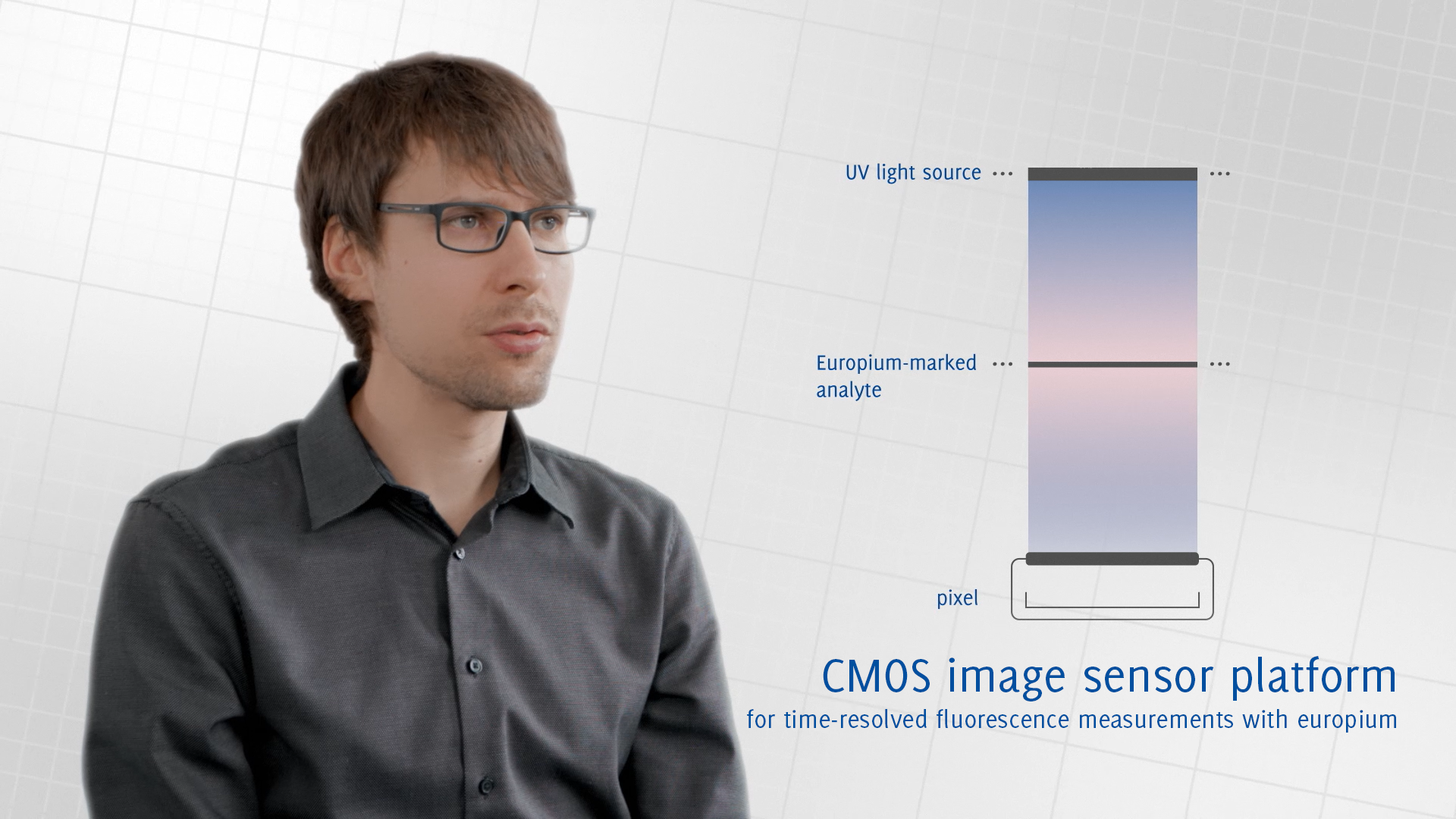
Classical fluorescence detection works with optical filters. Here, a UV light source illuminates an analyte that is marked with a fluorescent dye. In this case, this is europium which is excited by the UV radiation and produces a red fluorescent light. An optical filter then separates this light from the UV light and allows only the fluorescent light to reach the detector.
Time-resolved fluorescence detection
“Time-resolved fluorescence detection with lock-in pixels, as we have implemented it, does not require optical filters” explains Eric Schäfer, Head of Microelectronics at IMMS.“ Distinction between excitation and fluorescence light based on their colours is no longer possible - and we don't need it, because we use the different decay times of the light source and the fluorescent dye after switching off the light source.” While the excitation light decays within a few nanoseconds, the europium continues to glow for several 100 microseconds.
Lock-in-pixel principle
During excitation the pixel is deactivated, i.e. the charge carriers are dissipated. Afterwards, the pixel is activated and the charge carriers generated by the fluorescent light are collected in the pixel. Since the fluorescent light can be very little, it is useful to repeat this process and operate the pixel in the so-called lock-in mode. For this, the light source is pulsed and synchronously the pixel is always activated when the light fades away. This means that the fluorescence signal is being collected over several cycles and thus amplified. At the same time, the noise is reduced.
“We can adjust the number of cycles to the different light conditions: If we have a lot of light, a lot of fluorescent light, because we have a lot of analytes, for example, we can run fewer cycles. If we have relatively little, we can run more cycles” says Eric Schäfer. “This results in a very high dynamic range.” After the analyte has been excited several times and enough charge carriers have been collected, the sensor element is being read out and reset for the next measurement.
Imager platform for a wide range of applications in in-vitro diagnostics
“Since we have developed the sensor element as a pixel, we can use it to build entire image sensors, for example for optical imaging systems or for contact imaging” Eric Schäfer summarises.
The imager platform thus provides the basis for opening up a wide range of applications in in-vitro diagnostics. IMMS aims to use the platform to develop further application-specific time-resolved image sensors and the associated hardware and software modules.
Related content

Project
MEDIKIT
IMMS has developed a chip for mobile diagnostic systems for the early detection of diseases using time-resolved fluorescence measurements.

Event,
IID 2023
Industry Innovation Dialogue: New paths in microelectronics – diversification for more resilience
Event,
InnoCON Thüringen 2021
Die diesjährige InnoCON steht unter dem Motto „Mit der Thüringer Innovationsstrategie 2021 – 2027 die Herausforderungen unserer Zeit wie die digitale…
Event,
IEEE BioCAS 2021
Biomedical Circuits and Systems Conference (BioCAS): Restoring Vital Functions by Electronics – Achievements, Limitations, Opportunities, and…
Contact

Contact
Dipl.-Hdl. Dipl.-Des. Beate Hövelmans
Head of Corporate Communications
beate.hoevelmans(at)imms.de+49 (0) 3677 874 93 13
Beate Hövelmans is responsible for the text and image editorial work on this website, for the social media presence of IMMS on LinkedIn and YouTube, the annual reports, for press and media relations with regional and specialist media and other communication formats. She provides texts, photographs and video material for your reporting on IMMS, arranges contacts for interviews and is the contact person for events.